Radiation-free breast cancer screenings come closer to reality
An international research team has been leveraging the power of the German Research Network’s (DFN’s) X-WiN network to develop a more reliable, less invasive way to screen for breast cancer. Specifically, the team is focused on improving ultrasound technology to more accurately detect breast cancer in women who have so-called dense breasts, as traditional mammograms can miss the beginning of cancer or show false positives for these women.
“This is where we come in with ultrasound tomography to help,” said Dr. Nicole Ruiter, Department Head at the Karlsruhe Institute of Technology’s (KIT’s) Institute for Data Processing and Electronics. “With ultrasound imaging, we can avoid using radiation, meaning we could safely run scans on pregnant women and younger women. Further, our methods can screen at a higher frequency, and might deal better with accurately scanning women with dense breasts.”
Ruiter and her KIT colleagues are a part of a large European research project called QUSTom, which aims to revolutionize breast cancer screening. The two-and-a-half-year project aims to develop a workflow for fully 3D ultrasound tomography (USCT) breast cancer screenings. Headquartered at the Barcelona Supercomputing Centre (BSC) in Spain, QUSTom uses ultrasound imaging in concert with high-performance computing (HPC) in pursuit of its goals. For Ruiter and her KIT collaborators, access to the X-WiN networkis an essential piece of the technological puzzle needed to improve breast cancer screening accuracy and safety.
Imaging and simulation work together to advance the state-of-the-art
In the 1970s, researchers had begun experimenting with using ultrasound technology to scan for breast cancer. While the lab experiments showed promise, mammography became the preferred method for screening. Traditional ultrasound methods are primarily used only in situations where doctors are scanning soft tissue in the body, which in theory would work well for breast cancer screening. However, standard ultrasound imaging by itself can struggle to catch early warning signs for breast cancer—such as small calcium deposits forming—and are more prone to false positives or negatives in women with dense or fattier breasts.
In the last decade, researchers started actively designing methods to combine ultrasound technology with computed tomography—a method that normally uses x-rays that takes many images from different angles. Ruiter and other researchers in the field started focusing on USCT. Unlike traditional CT scans, USCT does not use x-rays for imaging, substituting ultrasound—albeit a different type.
Ruiter noted that the availability of fast data acquisition and management made USCT possible. “USCT is possible due to the availability of many parallel channels, doing fast data acquisition due to how many distributed ultrasound transducers we need, and then, of course, powerful computing and networking resources so that we can accurately reconstruct these images at high resolution.”
Stronger networks support advancements in medical technology
The QUSTom project involves six partners from Germany, Slovenia, Spain, and the United Kingdom. Further, ultrasound data of patients falls under protected medical data, meaning the project participants need to anonymize data before working with it.
“On the one hand, we must keep this information safe, and on the other hand, we are dealing with huge amounts of data that needs to be transported,” Ruiter said. “Our group is checking the data that is coming in and are creating starting models, so we are downloading it, checking for errors and accuracy, doing initial reconstructions, then reuploading analysis and the models. Another group does data preprocessing, and then that data is sent to a supercomputer in Barcelona. There is a huge need for high bandwidth, stable data movement and storage—we are collecting roughly 500 gigabytes of data per patient.”
The KIT team relies heavily on DFN’s X-WiN network to efficiently download and upload these massive datasets and transmit them to partners across Europe. DFN’s connections within Germany ensure that Ruiter can move data across German efficiently, and she is hopeful that connections forged through the GÉANT collaboration of Europe’s collection of national research and education networks (NRENs) continue raise speed and efficiency of moving data between institutions supported by different NRENs. Spain’s NREN, RedIRIS, connects BSC’s supercomputing resources to research organizations in Spain and internationally through the greater GÉANT collaboration, including Ruiter and her colleagues at KIT.
The QUSTom project has already successfully performed a feasibility study and continued work on improving prototype USCT devices. Moving forward, the project will continue to rely on high-resolution simulations supporting imaging being done at medical facilities. Project partners are also continuing to find ways to integrate their workflow into the computing cloud, ultimately striving for a “black box” technology that allows doctors to scan a patient, process the data quickly and securely, and give the patient accurate results as efficiently as possible.
This is an edited version of a story first published in DFN Mitteilungen publication. Read the original story.
Author: Eric Gedenk
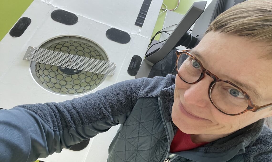
Featured image: As part of the EU-funded QUSTom project, Karlsruhe Institute of Technology Dr. Nicole Ruiter works with researchers across Europe to develop next-generation breast cancer screening devices. The team is actively creating prototypes for radiation-free breast cancer screening devices. Foto: KIT
For more information please contact our contributor(s):

